- Science
Early success with a procedure called a mitochondrial transplant offers a glimmer of hope for people fighting for survival after cardiac arrest, stroke, and more.
Published March 16, 2022
15 min read
If you saw six-year-old Avery in her dance class today, you’d never guess that she almost died from a heart defect. She underwent her first open heart surgery shortly after birth, and the procedure left much of her heart damaged. After two months in the hospital, she was deemed healthy enough to go home. But her mom, Jess Blias, rushed her back a few weeks later because Avery had “turned blue.” Her heart was only pumping at half its capacity, and she needed another surgery.
Doctors started preparing her for a heart transplant, but they noticed that during the brief moments when they disconnected her from ECMO, the machine that pumped her blood, in order to clean the tubes, her heart was functioning a bit better than they’d expected, suggesting the organ might be salvagable. That’s when Sitaram Emani, a cardiovascular surgeon and department head at Boston Children’s Hospital, approached Blias and offered to perform an experimental procedure to save her daughter’s life: a mitochondrial transplant.
This procedure involves collecting a patient’s mitochondria—tiny oval structures that provide cells with energy to function—and injecting them into damaged tissue. If it works as expected, the healthy mitochondria get absorbed into damaged cells and help them heal from the inside. With no other options for the infant Avery, “It was kind of like a Hail Mary,” Blias says. Amazingly, it worked. Avery’s heart pumped stronger each day, and she was able to go home. She’s had a total of six heart surgeries in as many years and still needs regular cardiological treatment, but if you saw her today, you’d never know anything was amiss.
Now scientists are betting that an infusion of mitochondria will jump-start the cellular processes required to heal damaged hearts, brains, and maybe even other organs in a way that drugs haven’t been able to. So far, the results from animal models and a few first-in-human trials like Avery’s have been promising. In the past few years new biotech companies have launched to harness the power of mitochondria for applications from wound healing to anti-aging.
There’s still a lot left to learn, and at this stage there is little government funding for this type of research. The Boston team, for instance, relies heavily on philanthropic donations. Michael Levitt, an associate professor of neurological surgery at the University of Washington, is working to transplant mitochondria into the brains of stroke patients, and he says his team has “no external funding whatsoever. This is all blood, sweat, and tears.”
Still, the scientists following this path are hopeful mitochondrial transplants will be a game-changer for treating a range of conditions, from wounds to strokes and heart attacks. “We’re so optimistic about what this could mean,” says Melanie Walker, a clinical professor of neurological surgery at the University of Washington and a colleague of Levitt’s.
Mitochondrial mayhem
Mitochondria are often described as “the powerhouses of the cell” because they manufacture a molecule known as adenosine triphosphate, or ATP. This molecule stores energy from the food you eat and uses it to fuel activities in different parts of your cells.
Scientists have long known that defective mitochondria can cause biological chaos. “Mitochondrial dysfunction is a universal driver of disease,” says Keshav Singh, a mitochondria researcher at the University of Alabama Birmingham who founded the Mitochondria Research and Medicine Society in the U.S. and India. Whether tissue damage is caused by disease or even space travel, faulty mitochondria are frequently involved.
Singh’s interest in mitochondria was piqued in the early 2000s when he stumbled on an article published in 1982. The authors had harvested mitochondria from antibiotic resistant cells and transferred them to normal mammalian cells that were still susceptible to the drugs. The vulnerable cells absorbed the mitochondria from the resistant cells—and became resistant themselves.
Singh’s takeaway: If these powerhouses could be transferred from one cell to another and retain their function, maybe they could be used to heal tissues with dysfunctional mitochondria.
Singh and his team started by developing genetically engineered mice whose cells produce fewer mitochondria. In 2018 they published a study showing that these mice had visible signs of premature aging, such as wrinkled skin and hair loss. When the scientists reactivated the gene to boost the number of mitochondria, the mice once again became hairy and taut-skinned. That worked demonstrated clearly that replenishing diminished mitochondria could restore tissue function in a living animal.
A convenient partnership and last resort
One of the most exciting potential applications is using mitochondria to mend broken hearts.
Physicians have limited options to repair tissue damage after ischemia, a blockage of blood flow to an organ. About 13 years ago James McCully, who studies mitochondria and heart surgery at Boston Children’s Hospital, noticed that after ischemia, no drugs could save the malfunctioning mitochondria in damaged heart tissue.
“We developed cardio-protective agents, but what we found all the time was that the mitochondria were damaged no matter what we did,” he says. Damaged mitochondria can swell or leak, starving the cell of energy and nutrients or sending signals that trigger apoptosis—programmed cell death. In his research, McCully had seen that the cardiac mitochondria had shrunk and gone from black to translucent. In turn, the heart couldn’t beat effectively.
“I had thought that perhaps there was another option,” he says. McCully developed a 30-minute procedure for isolating healthy mitochondria and then transplanted them into damaged tissues in petri dishes, and eventually in live mice and pigs.
As McCully was working in his lab, Emani was just down the street operating on newborn infants with heart defects. When he heard about McCully’s work, he wanted to collaborate. The surgeries Emani performs to repair a coronary artery are extremely risky, with a chance he could cut off blood flow to the baby’s heart. If that happens, tissue starts to die. The only solution was for Emani to place the infant on extracorporeal membrane oxygenation (ECMO)—a machine that pumps oxygenated blood through the infant’s body. Then all anyone could do was wait and hope that the heart tissue healed on its own. Many times, it didn’t.
Emani recruited McCully to find a better way to treat the infants on ECMO. “It was quite controversial, risky, courageous, foolish—call it what you will,” says Emani. “But we had no other choice. I mean, we knew that these patients would die without any additional help.”
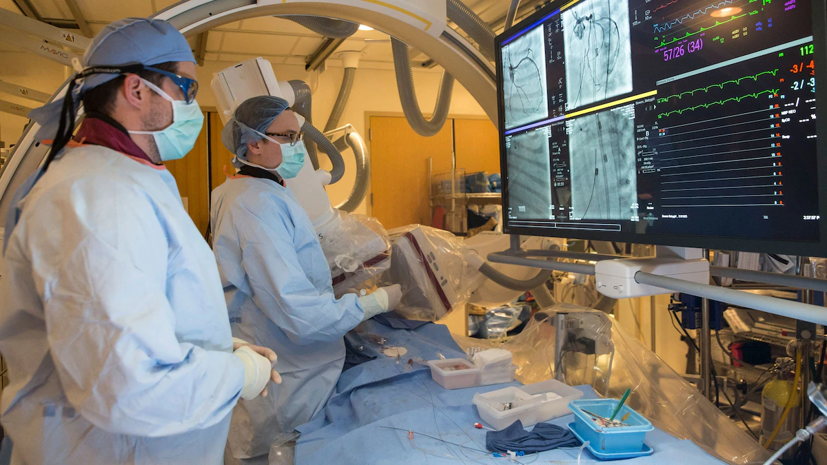
With chest open and heart exposed, the patient would lay on the table, still connected to the ECMO machine, while McCully would take a small tissue sample from the infant’s abdominal muscle. He would quickly harvest mitochondria from the muscle cells at a lab bench in the operating room. Then Emani would infuse about a billion of those mitochondria back into the patient’s heart through the coronary artery or via a direct injection close to the damaged region.
The team’s first patient in 2015 didn’t survive; the scientists believe they were too late. Time is of the essence here, because while the infusion can help rescue cells struggling with damaged mitochondria it can’t resurrect dead cells. Of the next 11 patients, one of which was Avery, eight survived. The New York Times touted the achievement in 2018.
They’ve only performed the procedure on three more infants since then, in part because surgical techniques have improved “so we’re not seeing this complication or issue as often,” says Emani. But based on their initial success, the researchers are now working with other hospitals to recruit pediatric patients for a clinical trial.
Jason Bazil, who studies mitochondrial injury in ischemia at Michigan State University, read the 2018 story and says he was skeptical at first. “I thought maybe the regenerative capacity of the young children was the primary reason for the recovery,” he says, rather than the infusion of mitochondria. But as he dug into McCully’s animal experiments, he grew more convinced the mitochondria were key.
At the Feinstein Institutes for Medical Research in New York, Lance Becker and Kei Hayashida believe that a mitochondrial transplant procedure like the one Avery had might improve the recovery of hundreds of thousands of people who experience cardiac arrest each year.
Becker’s goal is to rescue people hovering on the brink of death, and he is perhaps best known for pioneering techniques for therapeutic hypothermia—cooling down the body of a person experiencing cardiac arrest to slow tissue damage. Now, he’s hoping mitochondria transplants can have a similarly transformative impact on resuscitation medicine.
After inducing cardiac arrest and performing CPR in 33 rats, Hayashida used McCully’s technique to inject about a billion mitochondria into the leg veins of each rat. He found that 90 percent of the rodents survived the cardiac arrest, compared with only 40 percent of a control group that didn’t receive the mitochondria. The results are not yet published.
But Hayashida noticed something else during the experiments: the mitochondria might have been doing more than just healing the animals’ hearts. When the heart stops, patients can also suffer brain damage as blood flow to the head declines. Using special dyes, the team has tracked some of the transplanted mitochondria in the rats, which appear as glowing red dots, in the brain. It was “very surprising,” says Hayashida, that some of the mitochondria he’d injected into the femoral artery reached the brain, suggesting the injections may help heal both the brain and the heart.
Hayashida’s not the only one hoping mitochondria might help heal brains. As word spread about his work, McCully began training other researchers hoping to harness the powerhouse of the cell to heal tissue. The University of Washington’s Walker was one of the people who reached out to McCully to learn. She thought that if mitochondria could heal the heart after ischemia, why not the brain after an ischemic stroke?
A little bit of hope
Similar to ischemia in heart patients, a stroke cuts off blood flow to the brain. Even after the blockage is removed, substantial brain damage can result. “We’re basically plumbers when it comes to stroke. We can take the blockage out,” to restore blood flow, says the University of Washington’s Levitt. “But the damage to the brain is something we just don’t have a lot of control over.”
After learning how to collect mitochondria, Walker began experimenting with mouse models of stroke. Seeing those results “was really the ‘Holy crap, it works’ moment for me,” says Levitt, who bills himself as the skeptic to Walker’s optimism.
To translate the procedure to human patients, Walker reached out to Yasemin Sancak, a mitochondrial scientist in a separate lab at the University of Washington. Sancak says that she immediately thought, “this is crazy and is never going to work.” But after meeting the surgeons and reviewing their research, she was fascinated.
Like McCully in Boston, Sancak has taken on the role of isolating and purifying mitochondria from the patient while in the operating room with Walker. She collects the mitochondria, with the clock ticking, as the surgeons wait to inject them into their patient’s brain.
Together the team has treated three stroke patients with mitochondria transplants. So far, all they can say for sure is that the procedure was safe, but they suspect it also provided some benefit. Two patients “recovered reasonably well for the severity of stroke they had,” says Levitt. The third did not do as well, but the researchers suspect that’s because he’d had several strokes prior to the most recent one, worsening his prognosis.
The team doesn’t have objective measures for how effective the procedure is yet, but Walker says that there are some positive signs on their brain scans. Scans of stroke patients usually show a phenomenon called luxury perfusion, which are fuzzy, cloudy wisps that signal brain damage. The wisps generally don’t go away even after a person survives a stroke.
“We would never publish that; it would never hold up to scrutiny,” says Levitt. But a radiologist observed the scans with no knowledge of the team’s work with mitochondria and commented on how unusual it was that the luxury perfusion had diminished. That moment gave everyone a little bit of hope that the transplants really work. “It’s not just us,” he says.
Regulating regeneration
Thus far, mitochondrial transplants appear to be safe in both humans and lab animals. “There’s no inflammatory response,” says McCully. “We’ve had no adverse effects.” But Emani emphasizes that if the mitochondria were impure or broken, the outcome might be different, since those broken powerhouses can damage tissue instead of healing it.
Emani has been talking with the U.S. Food and Drug Administration about tightly controlling how these transplants are done. For years the FDA has been cracking down on a similar procedure, stem cell transplants, which were largely unregulated when they first emerged, leading to several high-profile adverse effects including blindness.
Because the transplanted mitochondria come from a patient’s own tissue and are injected during the same procedure as they are extracted, the FDA doesn’t require clinical trials and premarket approval. If the researchers were to take mitochondria from donor tissue, or harvest them from cells in the lab, the treatment would be regulated the same way as other drugs.
“We have to develop best practices,” Singh emphasizes.
There are also a lot of open questions, like whether injecting mitochondria into a leg vein will get enough of them to the heart, or if surgeons are better off opening the chest or skull and placing them directly next to damaged tissue.
Andrés Caicedo, a professor studying mitochondria at the San Francisco de Quito University in Ecuador, says he’s exploring the outcomes based on different sources of mitochondria. He hopes to use them for wound healing, and he suspects that mitochondria extracted from mature tissue like muscle might not be the best strategy. Because stem cells grow and differentiate more rapidly than muscle cells, he wonders if their mitochondria might be better suited for regrowing skin.
Another big question is just how many mitochondria are needed for each transplant. In his animal studies, McCully has found that gets the best results with a certain number of mitochondria based on the weight of the animal’s heart. But that ratio doesn’t apply to all tissues. If he’s injecting mitochondria into skeletal muscle, for example, he needs to inject more mitochondria per gram of muscle tissue.
Some researchers also say there’s good reason to try using donated mitochondria, or better yet, develop a cell line from which they can harvest and store ready-to-use mitochondria. It isn’t practical for most hospitals keep lab scientists like McCully and Sancak on call to isolate mitochondria at a moment’s notice. Having a source of transplantable mitochondria would not only speed up and standardize the procedure for emergencies, it might also allow them to treat patients with mitochondrial diseases.
“Ideally what we would have is a really good cellular source of mitochondria that grows in every hospital,” says Becker.
If nothing else, “I do think it’s time to start respecting mitochondrial transplantation,” says Bazil. “It’s time to zero in on this phenomenon and explain it, to try to save as many lives as we can.”

