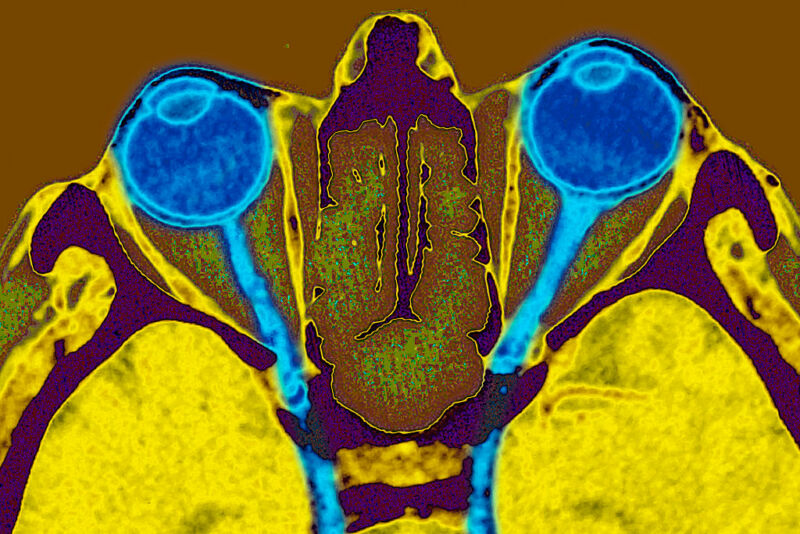
Can you see the light? — Reprogramming nerve cells and hardware implants both add to visual acuity.

Our visual system is complex, with photoreceptors that pick up incoming light and at least three types of neurons between those and the brain. Once in the brain, visual input is interpreted by multiple dedicated regions that build a scene out of small pieces of shape and motion. The outcome of that processing may be further interpreted by areas of the brain that handle things like reading or face recognition.
With all that complexity, lots of different things can go wrong. Accordingly, we’ll likely need multiple solutions if we want to try to correct these problems. So, it was nice to see the results this week of two very different approaches to tackling visual problems tested in experimental animals. One group manipulated biology to correct problems in the transmission of information between the eye and the brain, while another group used electronics to bypass the need for an eye entirely.
Refreshing nerves
One of the most exciting developments in tissue repair has been the recognition that we could convert many cell types into stem cells just by activating four specific genes. Unfortunately, activating those genes widely in mice kills them, as the genes also promote loss of normal cellular identity and uncontrolled division. A huge US-based collaboration suspected many of these problems were due to one of those four genes (called MYC), and so it focused on working with the remaining three. The first showed that activating these three in cells from older mice restored properties that were typical of younger cells without any loss of normal cell function.
From there, the researchers focused on their real target: the eye. Specifically, they focused on a population of cells that connect the back of the retina to the brain, called retinal ganglion cells. Failure of these cells, which can occur in diseases like glaucoma, results in the progressive loss of vision. At birth in mice, these cells are able to regrow connections between the eye and brain if those connections have been cut. But that ability is quickly lost.
So, the researchers damaged the optic nerve, then activated the three stem cell genes in the retinal ganglion cells. With the genes active, even in adult mice, the connection was restored. The same was true when they artificially induced glaucoma in these mice. Tests of their vision indicated that nearly half of the lost visual acuity was restored by this gene treatment. The same thing was true for the loss of visual acuity that comes with age, confirmed by comparing mice at three months of age to those roughly a year old.
All of this happened without the growth of new cells. Instead, the existing cells seemed to be able to repair or replace the damaged parts (called axons) that form the optic nerve. The researchers went on to show that this repair depends on changes in a type of chemical modification of DNA called methylation, which can alter the activity of lots of genes.
Bypassing the eye
The second study, done by four European researchers, focuses on events well downstream of the eye. Once signals get to the brain, they’re first interpreted by a region that has a one-to-one physical mapping with the retina. In other words, the geometry of neurons in the part of the brain that receives signals from the retina mirrors the layout of the retina itself. The researchers use this correspondence and some electronics to try to activate the visual system without involving the eye at all.
They rely on a set of electrodes called the Utah Array to make connections with the neurons in this area of the brain. The Utah Array doesn’t have that many electrodes—Elon Musk regularly makes reference to how many more his Neuralink hardware will have—but, since these are research animals, the researchers here simply implant a collection of Utah Arrays into primates. Implanting 16 individual electrodes into one brain is not something that’s going to receive approval if the subject is human, but it does what’s needed in a research context.
The researchers use these implants to not only wire up the area where visual signals reach the brain and are first interpreted; they also plug into the area where those interpretations go on to be processed. This helps them determine the right amount of current to inject into the brain in order to stimulate a small bit of the visual field without overwhelming it. These small injections create what are called “phosphenes”—what are perceived as small flashes of light. And, because of the geometry of this area of the brain, the researchers can control where the flashes of light appear in the visual field.
Generally, it worked. The primates would typically direct their eyes toward where they had perceived the flash of light to originate, even though nothing had actually happened at that location that would have registered on their eye. The monkeys were also trained to identify whether two dots were arranged vertically or horizontally, and they managed to do so when the “dots” were instead phosphenes generated by the electrodes. They weren’t as good as when physical dots were shown to them, but they definitely did much better than would be expected from a random choice.
More dramatically, the monkeys were also trained to recognize letters and did so even when the letter was created by an order set of current injections. In other words, the monkeys could recognize a pattern of phosphenes as depicting a letter—again, not as well as they managed when an actual letter was shown to them, but well above random chance.
Having visions
For starters, it’s important to be clear that both of these are just early attempts at figuring out what’s possible using research animals. We are nowhere close to treatments in humans. And it’s difficult to interpret just how much of a change in vision we’re producing via either technique, since we can’t ask lab animals what they see and have to rely on indirect tests of their visual abilities. And there’s a lot of potential safety issues here, especially for something that involves altering the activity of human genes.
That said, of the two, the gene-manipulation experiments are far more intriguing. We had already known that electrodes placed in the right area of the brain could produce visual artifacts when activated. To an extent, organizing the visual artifacts to convey information was a matter of engineering more than anything. But the apparent restoration of nerve function that’s lost due to age or injury is far more unexpected, as is the fact that it can be done using a relatively small genetic intervention. If it holds up to replication, it definitely seems to point toward applications that are far more expansive than vision.
Nature, 2020. DOI: 10.1038/s41586-020-2975-4; Science, 2020. DOI: 10.1126/science.abd7435 (About DOIs).
By John Timmer

